

Eye Anatomy
The eye is a complex organ that works much like a camera, focusing light rays and forming an image. On the surface of the eye is the cornea, a thin, spherical layer of tissue that provides a clear window for light to pass through. In a healthy eye, the cornea bends or refracts light rays so they focus precisely on the retina in the back of the eye.
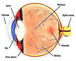
Beneath the cornea is the iris, the colored part of the eye we refer to when we say a person has brown or blue eyes. In the center of the iris is the pupil. The iris functions like a shutter, adjusting pupil size to control the amount of light entering the eye.
Located behind the iris is the lens, which works together with the cornea and vitreous to focus light. Like the lens in a camera, it adjusts light rays as vision shifts between nearby and distant objects in a process called accommodation.
Light then passes through the vitreous, the gelatinous substance that fills most of the eye and gives it its shape.
The back of the eye is lined with a thin layer of tissue containing millions of photoreceptor (light-sensitive) cells. This is the retina, where light rays focus into an upside-down image. In the center of the retina is the macula. Less than 1/4 of an inch in diameter, the macula is responsible for clear central vision. The retina converts the image into an electrical signal that travels down the optic nerve to the brain.
General Questions
What is an Ophthalmologist?
An ophthalmologist is a physician -- a medical doctor (MD) or doctor of osteopathy (DO) -- who specializes in the medical and surgical treatment of eye diseases or other conditions. Ophthalmologists also often provide routine vision care services such as prescribing eyeglasses and contact lenses. The training required to become an ophthalmologist is strenuous. It includes at least four years of medical school (after undergraduate college); a one-year internship in general medicine, usually at a hospital; and a three-year residency in ophthalmology at an accredited teaching program. Many ophthalmologists choose to complete one or two additional years of training in a fellowship, concentrating on a particular aspect of medical or surgical eye care.
What is an Optometrist?
An Optometrist (OD) is a doctor of Optometry that generally provides routine vision care services, prescribes eyeglasses and fits contact lenses. Currently Optometrists,diagnose and medically treat eye diseases. Optometrists may not perform any form of surgery.
What is an Optician?
An Optician is trained to dispense eyeglasses, but does not perform eye exams or treat any eye diseases. At the present time, opticians are not required to be licensed in most states.
What does board-certified mean?
Board certification means that an ophthalmologist has taken and passed rigorous examinations which cover all aspects of medical and surgical eye care. These examinations are voluntary. An ophthalmologist does not have to be board-certified to practice. The American Board of Ophthalmology (ABO) is the main certifying body for ophthalmologists in the United States. Medical specialty boards - including the ABO are accredited by an "umbrella" organization that sets standards for certifying physicians.
What does subspecialist mean?
For most eye problems, a general ophthalmologist or optometrist provides comprehensive care. While all ophthalmologists specialize in treatment of eye problems, some choose to concentrate in a more specific area. This is usually after completing a fellowship. Some sub-specialists focus on treatment of a particular disease, such as glaucoma. Others may specialize in a specific part of the eye like the retina or the cornea, or in a particular field such as pediatric care.
My eyes don't hurt and my vision is okay. Why should I have an eye exam?
Regular eye exams are an invaluable tool in maintaining your eyes’ health by detecting and preventing disease. Some diseases, such as glaucoma, often develop gradually without causing pain or vision loss. You may not notice anything wrong until significant and irreversible damage has been done. Early detection of any problems can allow for a choice of treatment options and a reduced risk of further harm.
What is Glaucoma?
One of the leading causes of blindness in the United States, glaucoma occurs when the pressure inside the eye rises high enough to damage the optic nerve. It cannot be prevented, and vision lost to it cannot be restored. The high eye pressure associated with glaucoma is caused by blockages in the eye’s fluid drains. No one knows yet why the blockages form. People at the greatest risk include those who are over the age of 40, diabetic, near-sighted, African-American, or who have a family history of glaucoma.
Does Glaucoma have any symptoms?
Glaucoma often develops over many years without causing pain – so you may not experience vision loss until the disease has progressed. Symptoms are occasionally present and should be taken as warning signs that glaucoma may be developing; these include blurred vision, loss of peripheral vision, halo effects around lights and painful or reddened eyes.
Is Glaucoma treatable?
Once diagnosed, glaucoma can be controlled. Treatments to lower pressure in the eye include non-surgical methods such as prescription eye drops and medications, laser therapy, and surgery.
What is Macular Degeneration?
Macular degeneration is the number-one cause of blindness in the United States. It occurs when the macula -- a part of the retina in the back of the eye that ensures that our vision is clear and sharp -- degrades or "degenerates," causing a progressive loss of vision.
Does Macular Degeneration have any symptoms?
Yes. They include:
- A gradual loss of ability to see objects clearly
- A gradual loss of color vision
- Distorted vision
- A dark or empty area appearing in the center of vision
Can Macular Degeneration be treated?
The "dry" form of macular degeneration has no treatment, but the "wet" form may be helped by
- Injections. Wet AMD can now be treated with new drugs that are injected into the eye (anti-VEGF therapy). Abnormally high levels of a specific growth factor occur in eyes with wet AMD and promote the growth of abnormal new blood vessels. This drug treatment blocks the effects of the growth factor.You will need multiple injections that may be given as often as monthly. The eye is numbed before each injection. After the injection, you will remain in the doctor's office for a while and your eye will be monitored. This drug treatment can help slow down vision loss from AMD and in some cases improve sight.
- Photodynamic therapy. A drug called verteporfin is injected into your arm. It travels throughout the body, including the new blood vessels in your eye. The drug tends to "stick" to the surface of new blood vessels. Next, a light is shined into your eye for about 90 seconds. The light activates the drug. The activated drug destroys the new blood vessels and leads to a slower rate of vision decline. Unlike laser surgery, this drug does not destroy surrounding healthy tissue. Because the drug is activated by light, you must avoid exposing your skin or eyes to direct sunlight or bright indoor light for five days after treatment.Photodynamic therapy is relatively painless. It takes about 20 minutes and can be performed in a doctor's office.Photodynamic therapy slows the rate of vision loss. It does not stop vision loss or restore vision in eyes already damaged by advanced AMD. Treatment results often are temporary. You may need to be treated again.
- Laser surgery. This procedure uses a laser to destroy the fragile, leaky blood vessels. A high energy beam of light is aimed directly onto the new blood vessels and destroys them, preventing further loss of vision. However, laser treatment may also destroy some surrounding healthy tissue and some vision. Only a small percentage of people with wet AMD can be treated with laser surgery. Laser surgery is more effective if the leaky blood vessels have developed away from the fovea, the central part of the macula. (See illustration at the beginning of this document.) Laser surgery is performed in a doctor's office or eye clinic.The risk of new blood vessels developing after laser treatment is high. Repeated treatments may be necessary. In some cases, vision loss may progress despite repeated treatments.
What is a Cataract? Who is at risk for developing them?
A cataract is a cloudy area in the normally clear lens in the front of the eye. Cataracts are caused by a chemical change of unknown origin in the eye, and cause blurred or distorted vision. People at risk for developing cataracts are over 55 years old, have had eye injuries or disease, have a family history of cataracts, smoke cigarettes or use certain medications.
Can Cataracts be prevented?
They cannot be prevented from forming, but early detection through regular eye exams can help maintain the clearest vision possible.
Are there symptoms associated with Cataracts?
There is no pain associated with the condition, but there are several symptoms that indicate failing vision due to cataracts. These include:
- Blurred/hazy vision
- Spots in front of the eye(s)
- Sensitivity to glare
- A feeling of “film” over the eye(s)
- A temporary improvement in near vision
How are Cataracts treated?
Vision loss from cataracts can often be corrected with prescription glasses and contact lenses. For people who are significantly affected by cataracts, replacement surgery may be the preferred method of treatment. During cataract replacement (the most common surgical procedure in the country), the lens is removed and replaced with an artificial one called an intraocular lens or IOL.
What is Diabetic Retinopathy?
Diabetic retinopathy is a complication of diabetes that weakens the blood vessels that supply nourishment to the retina (the light-sensitive lining in the back of the eye where vision is focused). When these weak vessels leak, swell or develop thin branches, vision loss occurs. In its advanced stages, the disease can cause blurred or cloudy vision, floaters and blind spots – and, eventually, blindness. This damage is irreversible.
Can Diabetic Retinopathy be prevented?
Yes. People with diabetes are most susceptible to developing it, but your risk is reduced if you follow your prescribed diet and medications, exercise regularly, control your blood pressure, and avoid alcohol and cigarettes. Regular eye exams are an integral part of making sure your eyes are healthy.
Can Diabetic Retinopathy be treated?
Although damage caused by diabetic retinopathy cannot be corrected, patients diagnosed with the condition can be treated to slow its progression and prevent further vision loss. Treatment modalities include laser and surgical procedures.
What is Dry Eye?
"Dry eye" is the term for when your eyes are insufficiently moisturized, either because they do not produce enough tears or because the tears have an improper chemical composition. It often occurs during the natural aging process, but it can also form as a result of eyelid or blinking problems, certain medications (antihistamines, oral contraceptives, antidepressants), climate (low humidity, wind, dust), injury, and various health problems (arthritis, Sjogren’s syndrome).
Symptoms include:
- Irritated, scratchy, dry, uncomfortable or red eyes
- A burning sensation or feeling of something foreign in your eyes
- Blurred vision
In addition to being uncomfortable, dry eye can damage eye tissue, scar the cornea and impair vision. Dry eye is not preventable, but it can be controlled before harm is done to your eyes.
How is Dry Eye treated?
Treatment for dry eye can take many forms. Non-surgical methods include blinking exercises, increasing humidity at home or work, and use of artificial tears or moisturizing ointment. If these methods fail, small plugs may be inserted in the corners of the eyes to limit tear drainage, or the drainage tubes in the eyes may be surgically closed.
Restasis is a new topical medication that causes improved natural tear formation.
What is Presbyopia?
Presbyopia is a vision condition in which the crystalline lens of your eye loses its flexibility, which makes it difficult for you to focus on close objects.Presbyopia may seem to occur suddenly, but the actual loss of flexibility takes place over a number of years. Presbyopia usually becomes noticeable in the early to mid-40s. Presbyopia is a natural part of the aging process of the eye. It is not a disease, and it cannot be prevented. Some signs of presbyopia include the tendency to hold reading materials at arm's length, blurred vision at normal reading distance and eye fatigue along with headaches when doing close work. A comprehensive optometric examination will include testing for presbyopia.